Scientists have long worked on mapping the brain. Image: Gerd Altmann, on Pixabay.
By Mariana Meneses
New technologies are giving neuroscience an unprecedented level of access to the brain. Numerous studies report on cases where Artificial Intelligence (AI) has allowed researchers to map brain regions and dynamics in detail, leading to a greater understanding of neural biology. At the same time, this growing access is also being used to simulate neural systems outside living organisms.
The company Eon Systems recently claimed to have produced an early version of a virtual embodied fly: a digital fruit fly in which a brain model is linked to a simulated body so that sensory inputs can help drive behavior. In the demo they describe, the system works in a loop, as the fly moves toward a food source, stops to groom when “virtual dust” builds up, and then resumes moving and feeding.
How fruit flies are helping us understand human brains | CBC British Columbia
Eon Systems explains that this is not a fully built digital animal from scratch, but an integration of existing scientific work: a detailed map of the fly brain’s wiring, a simplified model of neural activity, and a previously developed physics-based body simulation. In plain terms, the project tries to connect three things: what the fly senses, how a modeled brain processes that information, and how the body then moves in response. The company uses an everyday analogy: it is less like simulating every internal mechanism in a car engine and more like using the steering wheel, brake, and accelerator to guide the car’s overall behavior.
The system’s limitations are stressed in the company’s release: it models only a small subset of the fly’s senses and behaviors, relies on simplified neuron models rather than full biological detail, and leaves out major factors such as learning, internal states, hormones, and many layers of motor control. Also, some connections between brain activity and body movement are still approximated by hand rather than fully derived from biology.
As a result, the embodied fly should not be understood as proof that brain structure alone is enough to reproduce a real animal’s full behavior, but instead as an early research and demonstration platform, a first step for testing how a connectome-based brain model might be linked to a moving body.
In the video below, computer scientist Dr. Alex Wissner-Gross comments on the company’s achievement that “The Singularity has belonged exclusively to artificial minds, until now”.
The First Multi-Behavior Brain Upload |The Innermost Loop with Dr. Alex Wissner-Gross
One commentator on the breakthrough, writing on the platform The Medium, argues that the widely circulated narrative of the “first brain upload” significantly exaggerates what scientists actually achieved. The “brain upload” narrative emerged by combining several separate studies into a single storyline. One study produced the anatomical map, another built simplified neural simulations that could predict activity in specific sensory-motor pathways, and another created a realistic digital body of a fly using reinforcement learning rather than the connectome. A later preprint paper attempted to connect these elements by using the wiring diagram to guide a simulated controller, but even this system required additional training and does not reproduce the complete behavior of a living fly.
The commentary argues that presenting these distinct advances as a single breakthrough obscures the limits of what has actually been demonstrated and reflects exaggeration by the startup company more than current scientific reality. The real milestone, according to the writer, is the complete connectome of the fruit fly brain: a detailed wiring diagram of 139,255 neurons and about 54.5 million synapse connections, produced through a decade-long international collaboration combining machine learning with extensive human verification. In fact, the writer stresses that this dataset represents a major advance for neuroscience and is already enabling new discoveries about neural circuits.
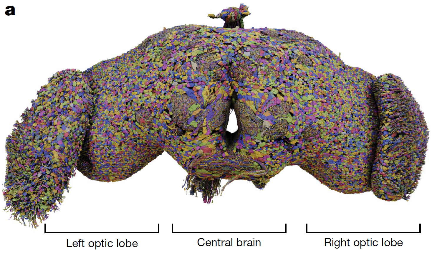
The first version of a full brain map of an adult fruit fly was earlier reported in a study entitled “Neuronal wiring diagram of an adult brain”, by Sven Dorkenwald and co-authors, published in the journal Nature (2024).
Rather than marking the end of the problem, these new simulations highlight how incomplete our understanding still is, one that new empirical research is continuing to deepen, particularly in the biological brain.
Reporting on a new study in this same field, Neuroscience News describes how researchers used artificial intelligence and MRI data from more than 50,000 individuals to map, for the first time, the genetic architecture of the corpus callosum, which is the bundle of nerve fibers that connects the brain’s left and right hemispheres. By automating the identification and measurement of this structure, the AI tool enabled large-scale analysis that would have previously taken years, revealing dozens of genetic regions that influence its size and thickness.
The article is entitled “The Genetic Architecture of the Human Corpus Callosum and its Subregions” was published in the journal Nature Communications in November 2025, by Ravi R. Bhatt, from the Imaging Genetics Center at the University of Southern California, and co-authors.
Neuroscience News explains that the findings show that different sets of genes shape distinct features of the corpus callosum and that many of these genes are active during prenatal development, when the brain’s fundamental wiring is established. Because this structure plays a central role in coordinating movement, integrating sensory information, and supporting higher-order cognition, variations in its development may help explain links to neurological and psychiatric conditions.
The study also identified genetic overlap between the corpus callosum, the cerebral cortex, and disorders such as ADHD and bipolar disorder, suggesting that shared biological pathways may underlie both brain structure and mental health risk. By making their AI tool openly available, the researchers aim to accelerate global efforts to better understand brain development and, ultimately, improve the diagnosis and treatment of related conditions.
Image: Gerd Altmann, on Pixabay.
In another example of the use of AI in brain research, Science Daily reported on a study where researchers used laser-based imaging and artificial intelligence to create the first comprehensive, dye-free molecular map of an Alzheimer patient’s brain. By combining hyperspectral Raman imaging (which is a technique that detects the chemical “fingerprints” of molecules) with machine learning, the team analyzed brain tissue in unprecedented detail, revealing patterns that traditional methods could not capture.
The article is entitled “Machine Learning-Enhanced Hyperspectral Raman Imaging for Label-Free Molecular Atlas of Alzheimer’s Brain”, and was published in the journal Biological and Medical Applications of Materials and Interfaces, in December 2025, by Ziyang Wang, from the Department of Electrical and Computer Engineering at Rice University, and co-authors.
Science Daily explains that the findings show that Alzheimer’s is not confined to the buildup of amyloid plaques, as traditionally believed. Instead, chemical changes are distributed unevenly across the brain, with some regions showing far greater disruption than others. This uneven pattern helps explain why symptoms develop gradually and why treatments targeting a single feature of the disease have had limited success.
Importantly, the study points to broader metabolic disruptions, particularly in memory-related regions such as the hippocampus and cortex. Changes in molecules linked to energy storage and cell structure, like glycogen and cholesterol, suggest that Alzheimer’s may be better understood as a whole-brain imbalance in energy and cellular function.
Alongside these advances, researchers are also using AI to better map white matter, the brain’s communication pathways that connect different regions. For example, MIT News has recently described how researchers developed an AI-powered tool capable of mapping distinct bundles of white matter fibers in the brainstem, an area that has historically been difficult to image in detail. Using diffusion MRI data, the system identifies and segments eight specific nerve pathways, enabling scientists to observe how signals related to essential functions like breathing, sleep, and consciousness travel through this critical region.
The study is entitled “Probabilistic mapping and automated segmentation of human brainstem white matter bundles”, and was published in the journal Proceedings of the National Academy of Science (PNAS), in February 2026, by Mark D. Olchanyi, from the Neuroscience Statistics Research Laboratory at the Massachusetts Institute of Technology, and co-authors.
The tool combines probabilistic mapping of fiber pathways with a convolutional neural network (CNN). CNNs are deep learning models designed to analyze structured grid-like data, such as images, for tasks like classification and object recognition. The CNN was trained on annotated brain scans, allowing it to reliably detect these bundles across different individuals and imaging conditions.
Once validated, the system was applied to patients with neurological conditions such as Alzheimer’s, Parkinson’s, multiple sclerosis, and traumatic brain injury, revealing distinct patterns of structural change in each case. These measurements, including bundle volume and the directionality of water diffusion along nerve fibers, provide new indicators of white matter integrity. The method also demonstrated the ability to track changes over time, including evidence of recovery in a patient emerging from a coma, where damaged nerve bundles gradually returned toward their original position.

Image: Gerd Altmann, on Pixabay.
In yet another development in this field, Quanta Magazine describes how researchers are using machine learning to transform brain mapping by organizing massive datasets of genetic activity from millions of individual cells. Instead of focusing on single cell types, the AI analyzes how different cells cluster together in space, much like identifying neighborhoods in a city, allowing scientists to build more meaningful maps of how the brain is structured.
The study is entitled “Data-driven fine-grained region discovery in the mouse brain with transformers” and was published in the journal Nature Communications in October 2025, by Alex J. Lee, from the Department of Neurology at the University of California, and co-authors.
Quanta Magazine explains that, by training an algorithm called CellTransformer on RNA data from millions of cells in mouse brains, the researchers were able to predict how cell types group together and generate high-resolution maps with up to 1,300 distinct subregions. These maps not only reproduced known brain structures but also revealed previously undetected subdivisions within larger regions, suggesting that areas once thought to be uniform may contain multiple specialized “neighborhoods” with different functions.
The approach highlights a shift in neuroscience where AI helps uncover patterns too complex for manual analysis. However, as these tools reveal increasingly detailed maps of the brain, they are also beginning to show the limits of what structure alone can explain.
Image: Mitrey, on Pixabay.
Neuroscience News has also recently featured how researchers have used advanced artificial intelligence models, including deep learning and convolutional neural networks, to test whether brain structure can predict navigation ability in healthy young adults. Analyzing MRI scans from 90 participants, the team looked for subtle patterns in regions such as the hippocampus, traditionally associated with spatial navigation, but found no measurable link between the size or shape of these structures and how well individuals navigated virtual environments.
The article is entitled “Deep learning approaches to map individual differences in macroscopic neural structure with variations in spatial navigation behavior” and was published in the journal Neuropsychologia in February 2026, by Ashish K. Sahoo, from the Department of Psychology at the University of Florida, and co-authors.
Neuroscience News explains that the findings challenge long-standing assumptions, partly shaped by studies of London taxi drivers who are required to memorize the city’s road networks, that better navigators have larger or differently structured brain regions. Even with tools capable of detecting complex patterns beyond simple measurements, AI failed to identify any “navigation signal” in brain anatomy. Performance was indistinguishable when comparing key regions like the hippocampus to control areas such as the thalamus, suggesting that macroscopic structure alone does not explain everyday navigation skills.
Instead, the results point to the importance of brain function and connectivity (i.e., how neurons interact) rather than overall size or volume. While AI has proven effective in identifying disease-related patterns, such as in Alzheimer’s, this study highlights its current limits in explaining normal cognitive variation. The research suggests that understanding abilities like navigation may require focusing on dynamic processes in the brain, rather than static anatomical features.
In this sense, what a system appears to reveal may depend not only on its true structure, but on how we choose to describe and measure it.
Phys.org describes how researchers have recently revisited one of the most basic concepts in physics, memory, and showed that in quantum systems memory depends on how the system is described. In classical physics, a process is considered memoryless if its future depends only on its present state. But in quantum mechanics, where measurement and information behave differently, this definition becomes more complex.
The article is entitled “Divisibility of Dynamical Maps: Schrödinger Versus Heisenberg Picture” and was published in the journal PRX Quantum in February 2026, by Federico Settimo, from the Department of Physics and Astronomy at the University of Turku, in Finland, and co-authors.
Phys.org explains that the researchers compared two fundamental ways of describing quantum systems: one that tracks how the system’s state evolves over time, and another that focuses on how observable quantities change. While both approaches yield the same experimental results, they reveal different aspects of memory. The study shows that some memory effects appear only when following the evolution of states, while others emerge only when tracking observables.
As a result, a quantum process can appear memoryless from one perspective while still retaining memory from another, challenging the idea that memory is a single, well-defined property. This finding suggests that quantum memory is richer and more nuanced than previously understood, with implications for quantum technologies, where managing noise and environmental interactions depends on accurately identifying and controlling these memory effects.

Sometimes we tend to see as binary what is not. Image: Gerd Altmann, on Pixabay.
Across these studies, a pattern emerges. As artificial intelligence allows us to map the brain in ever greater detail (its wiring, its chemistry, its cellular organization…) it also reveals a persistent gap between what we can describe and what we can explain. Structure, even when measured at scale and with precision, does not fully account for behavior, cognition, or individual differences. The brain is not only a physical object to be mapped, but a dynamic system whose properties depend on interactions, context, and time.
At a deeper level, this tension reflects a broader challenge that extends beyond neuroscience. As the quantum study suggests, what a system appears to reveal can depend on how it is described. More data and more detailed models do not simply uncover reality; they reshape the lens through which it is seen. The frontier, then, is not only about building better maps, but about developing better ways of interpreting them.
Craving more information? Check out these recommended TQR articles:
- Thinking in the Age of Machines: Global IQ Decline and the Rise of AI-Assisted Thinking
- Cleaning the Mirror: Increasing Concerns Over Data Quality, Distortion, and Decision-Making
- Not a Straight Line: What Ancient DNA Is Teaching Us About Migration, Contact, and Being Human
- Digital Sovereignty: Cutting Dependence on Dominant Tech Companies
We would appreciate your feedback on The Quantum Record and similar content.
Have we made any errors?
Please contact us at info@thequantumrecord.com so we can learn more and correct any unintended publication errors. Additionally, if you are an expert on this subject and would like to contribute to future content, please contact us. Our goal is to engage an ever-growing community of researchers to communicate and reflect on scientific and technological developments worldwide in plain language.