Image generated using Clipdrop.co.
By Saulo Silvestre and Mariana Meneses
This article was updated on September 20, 2023.
The use of three-dimensional imaging technology to study mummies has fundamentally transformed the field of archaeology.
This has offered researchers unparalleled insights into ancient civilizations while avoiding any harm to delicate artifacts.
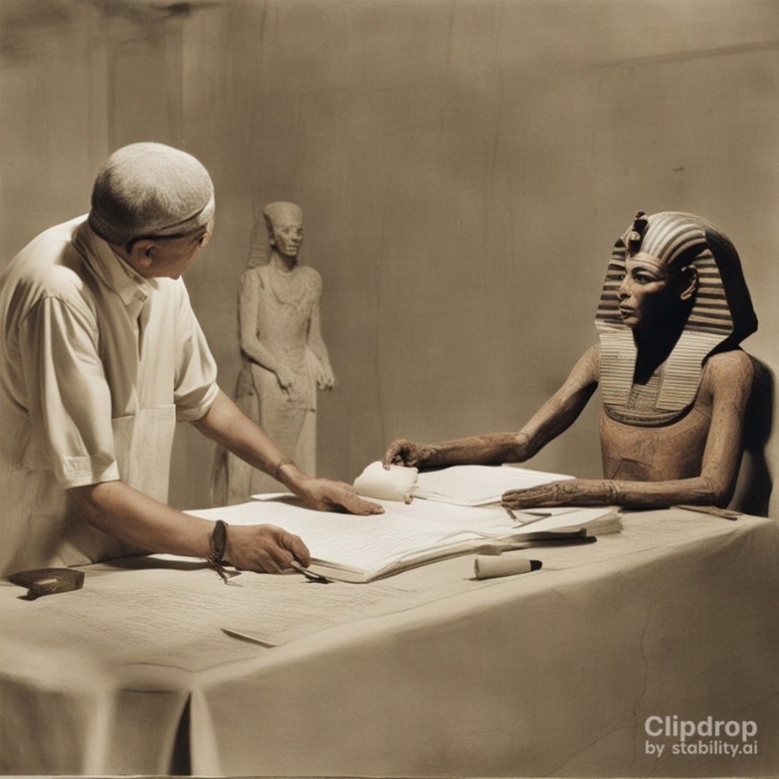
In two recent investigations, this remarkable technology was used to meticulously investigate the mummified remains of Egyptian pharaoh Amenhotep I and the enigmatic figure known as the “Golden Boy,” an ancient Egyptian mummy hailing from the Late Period (664 BC-332 BC). Through digital techniques to unravel and scan the mummies, researchers have uncovered fascinating new details of life over 2,000 years ago, without damaging the specimens so that future scientists might discover more. The mummy of Egyptian pharaoh Amenhotep I, one of the last remaining unwrapped royal Egyptian mummies, has been digitally uncovered and scanned in detail. Amenhotep I ruled Egypt from around 1525 to 1504 BC, during the New Kingdom era, and his mummy was found in 1881.
Due to its highly preserved wrapping and ornate face mask, it was left untouched, and its sarcophagus remained sealed… until recently.
“The New Kingdom, also referred to as the Egyptian Empire, is the period in ancient Egyptian history between the 16th and 11th century BC, covering the eighteenth, nineteenth, and twentieth dynasties of Egypt. Radiocarbon dating places the exact beginning of the New Kingdom between 1570 BC and 1544 BC. It was Egypt’s most prosperous time and marked the peak of its power.” (Wikipedia).

Illustration of pharaoh Amenhotep I. Richard Motel.
A paper published in December 2021 reported how researchers at the University of Cairo, Dr. Sahar Saleem and Dr. Zahi Hawass, used computerized tomography (CT) to peek inside this time capsule and map out the ancient king’s secrets.
Mummies provide an amazing source of knowledge.
Royal mummies of the New Kingdom, such as that of Amenhotep I, were the most well-preserved ancient human bodies ever found. Mummies like this can tell us about the appearance and health of ancient kings and queens, the mummification technologies used thousands of years ago, and cultural practices indicated by the funerary objects on the mummified bodies and in their tombs.
For centuries, Egyptologists have reconstructed many aspects of ancient Egyptian life and culture based on texts written on papyri, walls of tombs, and temples.
Although these inscriptions usually concern more formal aspects of Egyptian life, the mummies provide another story. Researchers often found another type of information on the preserved bodies that was not widely publicized. Mummies teach us about how these people ate, whether they had a healthy or poor diet, their health, age, genetic disorders, and sometimes even their cause of death.
Amenhotep I’s mummy had previously been examined using simple X-ray scans, but now scientists have managed to obtain a more complete and detailed picture of the internal body.
Using CT scans to examine the pharaoh’s bones, they found that he died at around 35 years of age and was about 1.69 meters tall. The new study allowed Dr. Saleem and Dr. Hawass to solve a long-standing mystery among Egyptologists. As it turns out, Amenhotep I was embalmed again 300 years after death by Egyptian priests, after his grave was robbed and his coffin was damaged. Interestingly, these priests may themselves have stolen precious jewels that were originally placed on the body and bandages of the ancient pharaoh.
The technique was so fruitful that it has resulted in further studies.
In new research, published in January 2023, Dr. Saleem conducted the scanning and three-dimensional-printing of the “Golden Boy” mummy, an ancient Egyptian mummy from the Late Period. The team unveiled insights into the mummy’s physical and social characteristics, such as bone structure, internal organs, and funerary objects. Furthermore, they used 3D-printing technology to create a replica of the mummy, which can be used for educational and research purposes without risking damage to the original artifact.
In 2021, when the first study was published, the scientists explained the reason to keep the mummy of Amenhotep I wrapped.
The priests did such good work of re-embalming the body that, more than 3,000 years after the pharaoh’s death, the preservation of his mummy astonishes archeologists. Of course, before modern imaging technology, it was not possible to study the contents of fully wrapped mummies. Back in 1821, when the first mummy autopsy was made, this was certainly not an option.
According to Dr. Daniel Antoine, curator of physical anthropology at the British Museum’s Institute for Bioarcheology, for most of the 19th century the only way to study mummies was to unwrap them, which usually damaged or destroyed these archeological treasures and often produced little useful knowledge. Moreover, mummies were not well protected in the nineteenth century, when unwrapping them was a popular form of entertainment among curious and wealthy Europeans. But times have changed, and so did our technology.
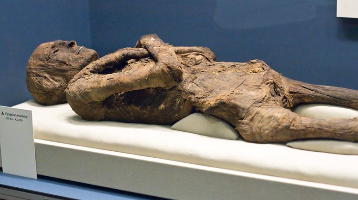
Egyptian mummy at Smithsonian Museum of Natural History, Washington. C. Watts.
Unwrapping mummies became increasingly uncommon during the 20th century when researchers began to use X-rays to study them.
In the 1960s, with the advent of portable X-ray machines, hundreds of mummies were scanned, including mummies on display in museums such as the British Museum, which holds the largest collection of mummies outside Egypt.
Thanks to this new technology, archeologists found evidence of broken bones, missing teeth, and amulets designed to protect from spirits, among many other findings. Nonetheless, these images were often blurred and did not allow for a very detailed examination. More recently, computerized tomography has been changing the way archeologists investigate such ancient Egyptian mysteries. This technology was invented in the 1970s and consists of X-rays from many angles rotating around the object under scrutiny. This technique allows scientists to collect a series of detailed two-dimensional images that are later combined to create three-dimensional pictures – in this case, of the body under the wrappings of the mummy.
In conversation with The Quantum Record, Dr. Saleem explained that “This application of using radiological imaging modalities for ancient objects and mummies is named Paleo-radiology. The birth of paleoradiology dates to the discovery of X-ray. The mysterious radiation (called X-ray) was discovered accidentally by the physicist Roentgen on November 8th, 1895. Before testing this unknown radiation on humans, it was tested on an ancient Egyptian mummy of a child and ancient Egyptian mummified cat; this was a few months later in mid-1896. For the first time, one could see what was inside the mummy without physically unwrapping it or dissecting, hence destroy this precious artifact for a mummified human.”
Today’s state-of-the-art CT scanners can have two sources of X-rays of different wavelengths, which significantly improves the clarity of the images.
Modern graphics software allows researchers to create virtual models of the mummies, which can be comprehensively explored without any harm to the body. According to Dr. Antoine, with this technology researchers can now virtually lift bandages from the skin, separate skin from muscles, lift muscles to reveal the bones, and even look inside an artery of the body inside fully wrapped mummies.
Dr. Saleem explained that she “used CT to scan hundreds of mummies as the main contributor to the Egyptian Mummy Project of the Egyptian Ministry of Antiquities, and [that they] plan to scan even more in the near future.” In her view, “Paleoradiology is expected to have rapid growth in the future as a specialist since it can work with cutting-edge imaging technologies as well as software for reconstruction and graphics. Dual energy CT can give us more precise information regarding mummification, thus I anticipate that it will be useful for imaging mummies in the future.”
There are still occasions in which some scientists believe it is worth unwrapping mummies.
Although imaging and virtual reconstruction are considered the gold standard for studying mummies without damaging them, more invasive methods may still be needed to produce information not available by other means. For instance, extracting and examining small pieces of soft tissue under the microscope provides scientists with better data on the presence of parasites and pathogens.
Another tool now used by researchers is DNA analysis, although today’s DNA technologies still face many challenges in the study of mummies.
For instance, the high temperature and moisture in the burial chambers, as well as the substances applied to the bodies during the embalming process can degrade the genetic material over time. Scientists are, however, excited about the next series of advances in DNA sequencing technology. They believe it will make it easier to reconstruct DNA from tiny fragments to help us unlock another set of mysteries, such as the origins and ethnic evolution of ancient Egyptians.
A group of researchers just published a paper on the comprehensive genome of the Tyrolean Iceman, an exceptionally well-preserved mummy discovered in the Italian Alps in 1991.
Through a thorough analysis of his DNA, they have made groundbreaking revelations about his ancestry and physical characteristics. The study suggests that there was significant migration in Europe during the Neolithic era, much more than ever thought. Furthermore, the research uncovered details such as his brown eyes, lactose intolerance, and heightened susceptibility to coronary heart disease. This study offers invaluable insights into the genetic history of Europe and provides a glimpse into the life of the Tyrolean Iceman.
We are now uncovering secrets from thousands of years ago that previous generations were unable to obtain – at least, to the best of our current knowledge.
Some argue, though, that if there are some aspects of mummies that we cannot study today without causing any damage, we should wait for just a little longer for our methods to evolve. Technology has developed extensively over the past few decades and will likely continue to advance rapidly. Egyptologists and other scientists of the future will only advance humanity’s knowledge of the archeology and human history embedded in mummies if we keep them protected long enough. Protecting these treasures might be worth the wait.
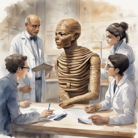
Because some fought to preserve mummies in the past, new methods now allow the extraction of more information than previously available. We should be aware of how our immediate interests may hamper our future ability to remember and learn from the past. Image generated using Clipdrop.co.
Keen to explore further? Dive into our vast TQR article collection exploring captivating discussions on thought-provoking topics such as:
- Ancient Technologies
- Dark Energy Explorers: A Citizen Science Project Looks Over 9 Billion Years Into the Past
- Magic Tricks for Birds Reveal a New Understanding of Human Minds
- Life Is Defined by Biology, Not Physics
- Amazing Evolutionary Leaps: Observing the Rapid Evolution of Single Cell Organisms into Complex Lifeforms
*** We use free AI software to generate text-to-image graphics to help convey abstract ideas in our articles. We give credit to the software each case, but we want to emphasize that we are not promoting any of them.